
Corneal Cross-Linking (CXL) is a minimally invasive procedure designed to stop or slow the progression of keratoconus and other conditions that cause the cornea to thin and weaken. By reinforcing the natural structure of the cornea, this treatment helps preserve vision and prevent the need for a future corneal transplant. At CarlinVision, we use the latest FDA-approved corneal cross-linking technology to ensure optimal safety, precision, and long-term results. Our experienced ophthalmologists specialize in personalized, evidence-based eye care that helps patients maintain stable vision for years to come.
What Is Corneal Cross-Linking?
Corneal cross-linking (CXL) is a specialized keratoconus treatment that strengthens the natural collagen fibers within the corneal tissue. The procedure combines riboflavin eye drops (a form of vitamin B2) with controlled ultraviolet (UV) light exposure to create new collagen bonds in the cornea. This photochemical reaction increases the strength and stability of the corneal structure, preventing further thinning or bulging.
During the corneal cross linking procedure, the outer layer of the cornea (epithelium) may be gently removed or loosened to allow better absorption of the riboflavin drops, depending on the specific technique used. Once the cornea is saturated, a focused beam of UV light activates the riboflavin, encouraging the formation of additional collagen cross links that reinforce the corneal framework.
This FDA-approved treatment has been clinically proven to halt or significantly slow the progression of progressive keratoconus and corneal ectasia after LASIK. By stabilizing corneal shape, patients often experience improved vision and reduced dependence on glasses or contact lenses.
Who Is a Candidate?
Corneal cross linking is recommended for patients diagnosed with keratoconus or post-LASIK ectasia, both of which cause the cornea to thin and bulge forward, resulting in distorted or blurred vision. Candidates include those with:
- Progressive keratoconus, where the cornea continues to weaken and change shape
- Corneal ectasia after laser vision correction surgery
- Difficulty achieving clear vision even with hard contact lenses or glasses
- Noticeable changes in corneal shape or curvature measurements
- No significant corneal scarring or other advanced damage
CXL is most effective in the early to moderate stages of keratoconus, before significant vision loss or corneal irregularities develop. For patients in severe cases, additional treatments such as specialty contact lenses or a corneal transplant may be necessary.
The Procedure
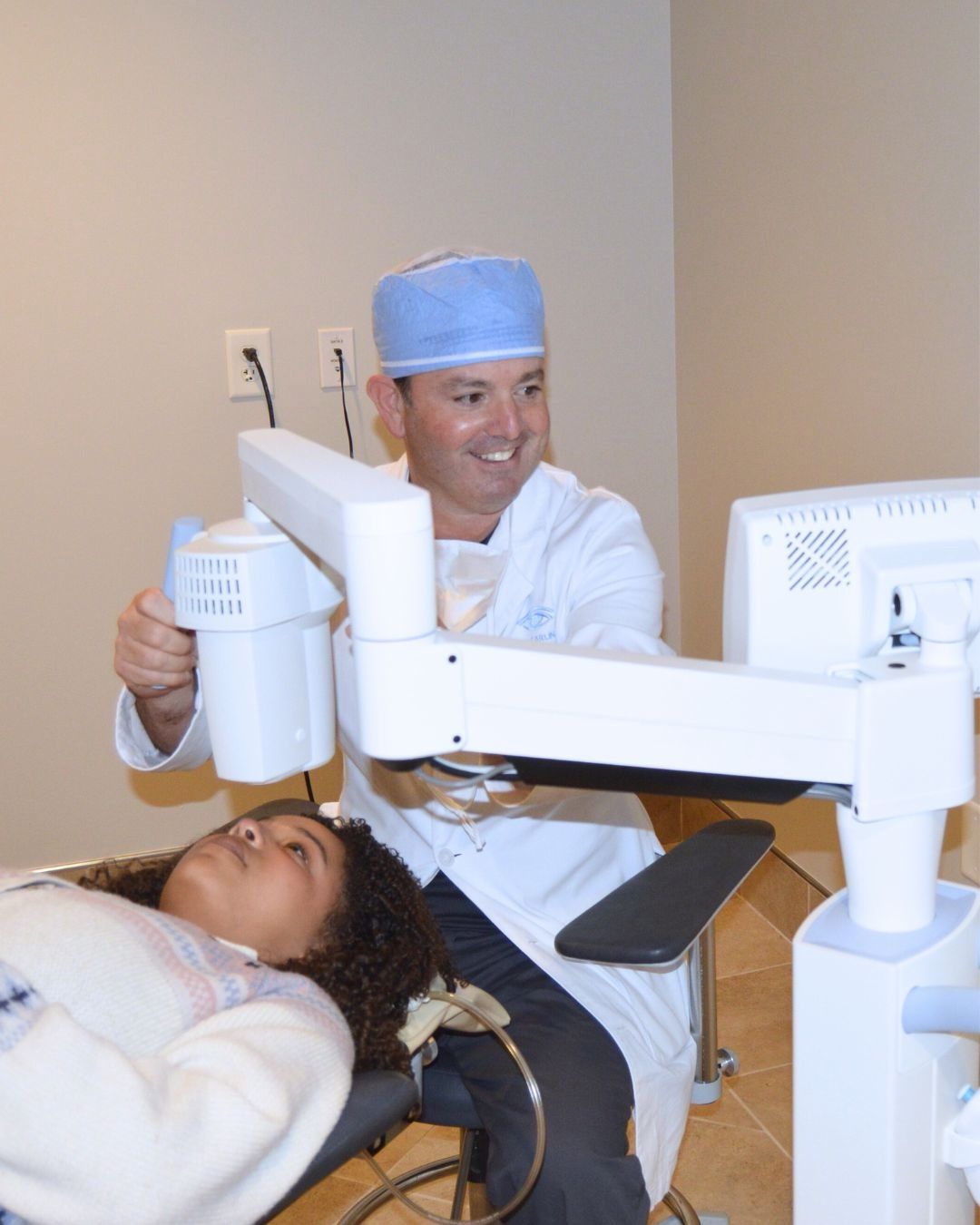
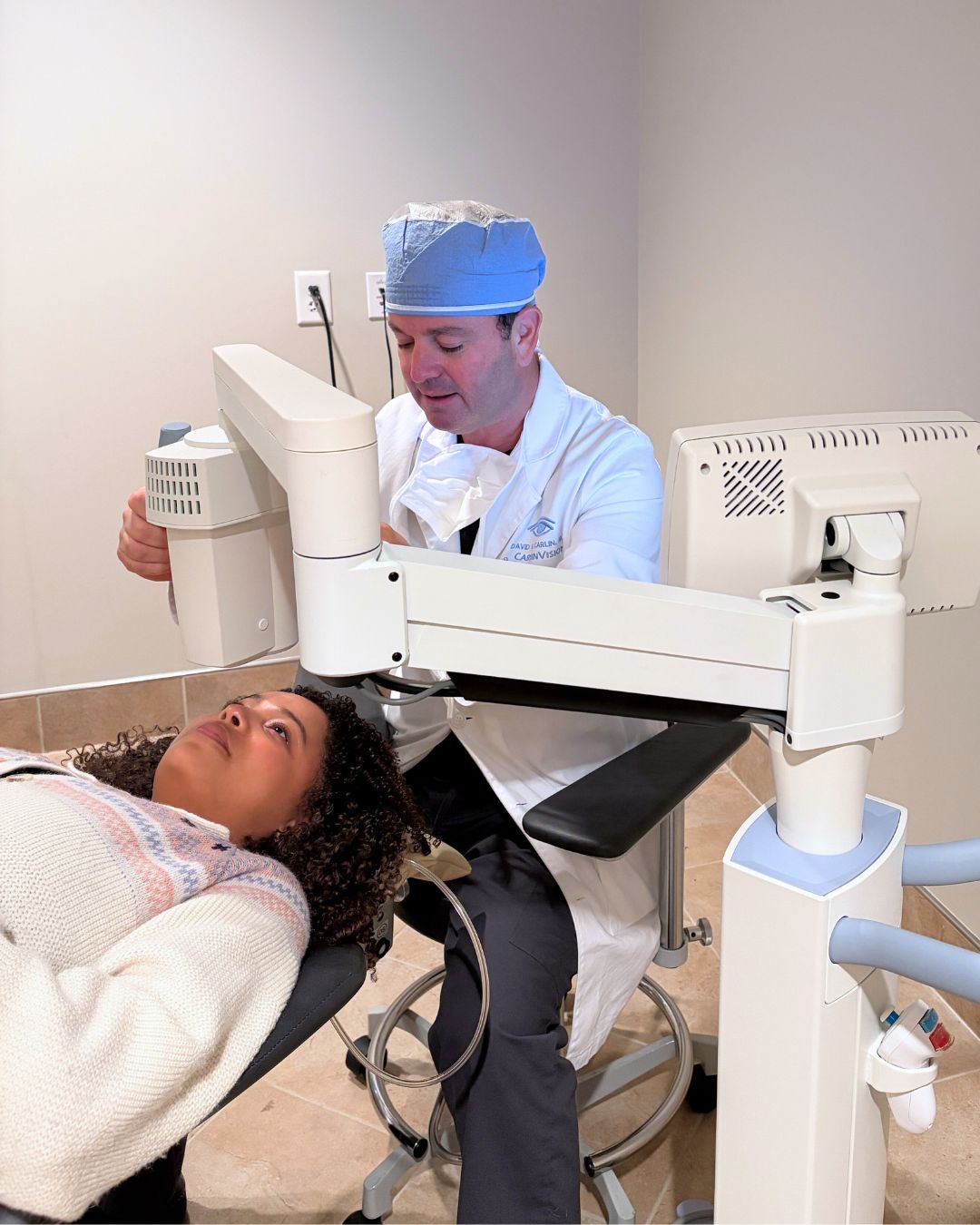
Corneal cross-linking surgery is an outpatient procedure that typically lasts about an hour. Patients remain awake during treatment, and anesthetic (numbing) drops are applied to ensure comfort throughout. The typical steps in each treatment plan include:
- Preparation: Your ophthalmologist cleans the surface of the eye and applies numbing drops. In the “epithelium-off” technique, the outer corneal layer is gently removed to help the riboflavin penetrate the corneal tissue.
- Riboflavin Application: Specialized riboflavin eye drops are applied to the cornea over several minutes to allow full saturation.
- UV Light Exposure: A controlled ultraviolet light (UV-A) source is directed onto the cornea for approximately 30 minutes. The combination of riboflavin and UV light forms new collagen bonds, strengthening corneal fibers and stabilizing shape.
- Completion: After treatment, a bandage contact lens is placed over the eye to protect the surface and aid healing.
Recovery and Results
Most patients experience mild discomfort, tearing, or light sensitivity in the first few days after CXL. These are normal signs of healing. The bandage contact lens typically remains in place for several days while the corneal epithelium regenerates. Our team may prescribe antibiotic and steroid drops to prevent infection and reduce inflammation.
The initial recovery time is about a week, during which you may experience some blurred vision or dry eyes. Vision gradually improves as the cornea stabilizes over the following weeks to months. It’s important to attend all scheduled follow-up visits so your doctor can monitor healing and corneal shape changes.
While corneal cross-linking does not reverse existing damage, it effectively halts disease progression in the majority of patients with keratoconus. Many patients notice improved corrected vision (with glasses or contact lenses) once the cornea has fully healed and reshaped.
Benefits of Corneal Cross-Linking
- Stops keratoconus progression
- Preserves vision
- Prevents corneal transplant
- Improves corneal strength
- Long-term stability
- Safe and proven
Why Choose CarlinVision?
At CarlinVision, your eye health and comfort are our top priorities. Our ophthalmologists are leaders in the diagnosis and management of keratoconus, using advanced imaging and FDA-approved CXL systems to deliver precise, effective outcomes.
Patients trust CarlinVision because our board-certified eye surgeons bring extensive experience performing corneal collagen cross-linking and other vision-saving procedures. We combine state-of-the-art diagnostic technology with highly personalized treatment planning to ensure the best possible results for each patient. From early diagnosis through post-procedure follow-up, our team provides comprehensive, compassionate care tailored to your individual needs. We focus not only on halting disease progression but also on protecting and enhancing your vision for the long term.
Schedule a Corneal Cross-Linking Consultation
If you’ve been diagnosed with keratoconus or are experiencing vision problems such as distorted or blurred vision, don’t wait to seek help; contact CarlinVision to schedule a corneal cross-linking consultation with one of our experienced ophthalmologists. We’ll perform a detailed examination to determine if CXL is the right treatment option for you and create a customized plan to protect and stabilize your vision. Call Mandy at 770.979.2020 x226 or email LASIK@carlinvision.com


Dr. Carlin is a highly experienced, board-certified ophthalmologist and Diplomate of the American Board of Ophthalmology with more than five decades of experience in vision care and surgical innovation. A graduate of the University of Illinois College of Medicine, he completed his ophthalmology residency at the University of Michigan before serving in the United States Navy in Key West, Florida. In 1977, he founded CarlinVision and has since built a longstanding reputation for excellence in LASIK, cataract surgery, refractive procedures, and cosmetic eyelid surgery. Throughout his career, Dr. Carlin has completed more than 11,000 procedures and remains at the forefront of advanced laser and refractive technologies. He is a Fellow of the American Academy of Ophthalmology and an active member of multiple leading professional organizations, reflecting his enduring commitment to clinical excellence and patient care.
Contact Us
Enter your contact info and a member of
our staff will contact you promptly.